|
MUSCLE
|

|
Abstract: Low back pain (LBP) is well known on earth, but is also known in space flight. This is remarkable because on earth LBP is mostly ascribed to heavy spinal load. A theory was developed which described the effect of flexing the lumbar spine in situations with negligible compressive load, i.e. non-weightbearing. This is in contrast with literature which ascribes LBP to excessive spinal load. According to our biomechanical model, verified by experiments, strain on the iliolumbar ligaments - which run from the lowest lumbar vertebra laterally to the hip bones - increases with backward tilt of the pelvis combined with forward flexion of the spine. A pain response can be the result from stress at the ligamento osseous junction of the iliolumbar ligament at the ilium and strain on the innervated iliolumbar ligament. This is what astronauts/cosmonauts may experience due to loss of lumbar curvature. Protection against prolonged strain can be provided by a deep muscle corset formed by the sacral part of the multifidus and the transversus abdominis muscles which act in co-contraction and have a separate part in the cortex. In microgravity this protection may be weakened due to muscle atrophy and neuro-muscular remodelling. If this phenomenon can be demonstrated, countermeasures can be developed. Details of the development in time of low back pain during flight are not known. Therefore a questionnaire will be completed during flight every day.
Address corresponding
author:
Chris J. Snijders* & Carolyn
A. Richardson**: Erasmus MC, University Medical Center Rotterdam Dept.
of Biomedical Physics and Technology P.O. Box 1738 3000 DR Rotterdam The Netherlands
tel: +31 10 408 73 68 fax: +31 10 408 94 63 e-mail: c.snijders@erasmusmc.nl.**The
University of Queensland, Faculty of Health Sciences Department of Physiotherapy
St. Lucia Q4072, Queensland Australia tel. +61 7 3365 2209 fax +61 7 3365 2775
e-mail: c.richardson@shrs.uq.edu.au.
Co-Investigators (all TNO-Human Factors):
Julie A. Hides**, Annelies
L. Pool-Goudzwaard*
Introduction: Musculoskeletal disorders form the single most expensive disease in society. Low back pain (LBP) forms the greatest contribution and despite ongoing research the incidence of LBP and the costs of its influence on society continue to rise. For 90% of the patients, the aetiology of the pain remains obscure. The one-year prevalence is 36%, which figure increases. Based on epidemiologic studies numerous causes of LBP have been suggested. Various pathological lesions are possible in the different structures of the lumbo-pelvic region, but even with modern technology the pain source is usually very difficult to diagnose accurately. For this reason there is a plethora of treatments for this condition, which have been developed by various medical disciplines. The challenge has been put forward, that treatments for LBP must have scientific evidence of their effectiveness. Also reported in literature is that up to 72% of shuttle crewmembers experience some form of LBP during flight and 28% of astronauts report moderate to severe LBP (Thornton, 1987; Fuchs, 1980). This is remarkable because on earth LBP is mostly ascribed to heavy spinal load with detrimental effects on the intervertebral discs (Wilke, 1999). Still this concept does not explain the causes of 80-90% of LBP cases. Therefore at Erasmus MC a different approach was chosen which a) included the sacroiliac joints, b) added the iliolumbar ligament as one of the most important structures at risk, c) related low back pain with pelvic floor disorders and d) included the non-weightbearing situation.
State of the knowledge on LBP
Biomechanical model
We developed a biomechanical model on the transfer of gravity load through
the lumbar spine and pelvis, with special attention for the sacroiliac joints.
The model predicts a significant stability effect of deep muscle forces (Snijders,
1995, 1998, 2002; Richardson, 2002), which is shown to be effective in the treatment
of low back pain (Richardson, 1999). This deep muscle corset was not included
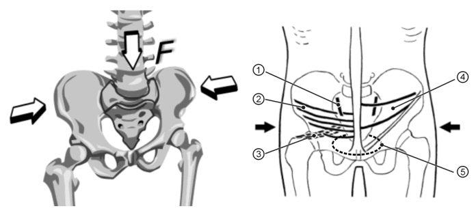
in earlier space studies. The above model is on the frontal plane (see Figure
1). The model developed on the sagittal plane mechanics explains iliolumbar
ligament tension in flexion and ease of strain by erector spinae and multifidus
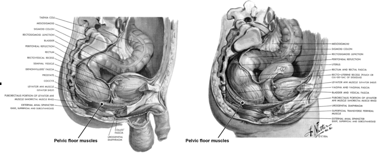
muscle force. The agonist-antagonist model about the sacroiliac joints (Snijders,
1993) relates back muscle dysfunction with pelvic floor muscle dysfunction (see
Figure 2).
 Figure 1: Transversely oriented muscles press the sacrum between the hip bones. This deep muscle corset for lumbopelvic stability is active with gravity load. Sacroiliac joint (1). Muscles: transverse abdominal (2), piriformis (3), internal abdominal oblique (4), pelvic floor (5). |
 Figure 2: The sacroiliac joints (upper left) link pelvic floor muscle dysfunction with back muscle dysfunction. |
Hypothesis: The deep sacral part of the multifidus and transversus abdominis muscles will show disproportional atrophy, neuroplasticity and morphological changes in flight, resulting in gradual development of low back pain in the course of ten days.
Methods: This is an in-flight study. The main scientific objective of the experiment is to assess astronaut/cosmonaut deep muscle corset atrophy in response to microgravity exposure. We propose to record the (expected) development of low back pain by means of a questionnaire. Type and intensity of pain recorded after every day will be related to the data on LBP development on earth and on the physiological process of muscle adaptation as obtained from the Berlin Bed Rest Study. In this study we measured changes in morphology and muscle volumes by means of MRI in the lower back and pelvic region and we used the questionnaire on low back pain.
Benefits field of research: Based on the results of the questionnaire countermeasures can be developed and implemented. This will involve ground studies and in flight experiments. Future research will involve selection and development of hardware. Ideas exist about special instrumentation to be developed for microgravity application as well as on earth.
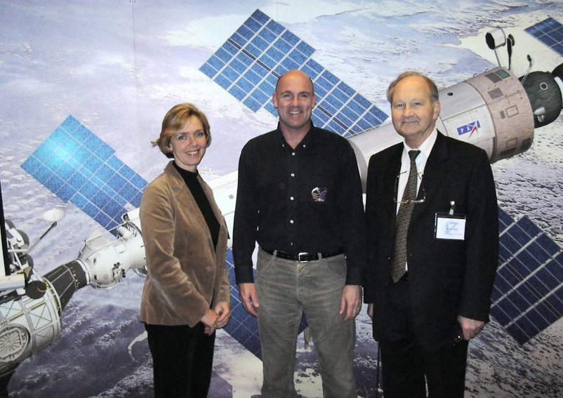
 Figure 3: On this picture in the middle the Dutch astronaut André Kuipers MD, next to him prof.dr. Chris Snijders and dr. Annelies Pool-Goudzwaard. The picture was taken at the Familiarisation Meeting at ESTEC, in the presence of other Russian and European astronauts. (photo: Antonio Torres, ESA/ESTEC, Noordwijk) |
Related papers: