Chapter 8
Otoconial alterations after embryonal development in hypergravity
HNPM Sondag1, HAA de Jong1, J van Marle2, B Willekens3, WJ Oosterveld1
1Vestibular department, ENT; 2Department of Electron Microscopy, Academic Medical Center, University of Amsterdam, The Netherlands; 3 Interuniversity Institute for Ophthalmology, Academic Medical Center, The Netherlands
Published in Brain Research Bulletin 40:000-000, 1996
Abstract
The relation between prolonged hypergravity and structural adaptation of otoconia was studied in hamsters (n=56). Three groups of hamsters (n=27), were conceived and born in a centrifuge: group 1 (n=10) 1 month under 2.5 G, group 2 (n=9) 5 months under 2.5 G and 4 months under 1 G, group 3 (n=8) 1 month under 2.5 G and 8 months under 1 G. Control hamsters (n=29), were conceived and born under 1 G (1 month old, n=7; 9 months old, n=22). Histological study of the otoconial layers (energy dispersive X-ray element analysis and scanning electron microscopy) showed similar calcium content, size and shape in utricular and saccular otoconia in all groups. Different were the utricular otoconial size classes, large, medium-sized and small. The area with small otoconia increased in group 1 (p=0.002). In group 2, the large otoconial area decreased (p<0.001) and the medium-sized one increased (p<0.001). In group 3, the large otoconial area decreased (p=0.003) and the medium-sized one increased (p=0.007). For age-related effects we found group 1 with an increased area of large otoconia (p=0.001) and a decreased medium-sized one compared to groups 2 (p<0.001) and 3 (p=0.02). Conclusion: hypergravity during formation of otoconia does not affect calcium content, size or shape, but changes the relative size of the areas with large, medium-sized or small otoconia and the development of these areas. This resulted in a structural adaptation to hypergravity.
Introduction
Correct spatial orientation requires perception
of gravity. The vestibular end-organs, the utricle and saccule, detect changes
in gravity. The otoconia on the maculae, as weight-lending structures, change
if exposed to prolonged altered gravity (Lim et al. 1974; Vinnikov et al. 1979;
Ross et al. 1985; Ross, 1987; Krasnov, 1991; Pedrozo and Wiederhold, 1994; Hara
et al. 1995. These structural changes can lead to inadequate vestibular sensory
information after return to normal gravity and are responsible for a disturbed
behaviour related to spatial orientation.
The otoconial genesis takes place during pre- and post-natal development (Ross,
1979; Kawamata and Igarashi, 1993; Kido et al. 1993). Especially in these periods,
otoconial changes can be expected during hypergravity. Experiments directed
to the effect of HG on the otoconia are sometimes conflicting. Krasnov (1991)
found no large otoconia in the peripheral part of the utricular patch in rats
developed under 2 G, whereas Hara et al. (1995) did find giant otoconia in the
same area in chick embryos developed under hypergravity. In hamsters, subjected
for 6 months to hypergravity after weaning, no differences were found in calcium
content, size, shape and distribution of the otoconia (Sondag et al. 1995).
The present study was performed to study the effect of hypergravity on the otoconia
in hamsters and in particular whether indeed otoconia structurally adapt to
hypergravity in hamsters conceived, born and raised in a centrifuge under 2.5
G for different periods. We assessed the calcium content, the shape and the
size of the otoconia on the saccular macula and the utricular macula as well
as the distribution of large, medium-sized and small otoconia on the utricular
macula.
Methods
Male golden hamsters (Mesocricetus auratus),
conceived and born under 2.5 G in a centrifuge (hypergravity or HG hamsters),
remained there for different periods of time. We studied 3 HG groups and compared
these to groups of controls, hamsters of the same age but conceived and living
under normal gravity conditions (CON hamsters): HG group 1 (n = 10) stayed for
1 month in the centrifuge; HG group 2 (n=9) stayed for 5 months in the centrifuge
and 4 months under 1 G; HG group 3 (n=8) stayed for 1 month in the centrifuge
and 8 months under 1 G. For HG group 1 we used control 1-month control hamsters
(n=7), for group 2 and 3 we used the same 9-months controls (n=22). Food and
water were ad libitum. The hamsters were killed after 1 month (n=17) or 9 months
(n=39). The experiments were performed in accordance with the recommendations
provided in a special license as required by the Dutch Law on the Care and the
Use of Animals in Scientific Research.
The animal centrifuge consisted of a centrally placed 3.5. kW DC motor drive
and 2 horizontally mounted arms with a length of 1.15 m. Each arm was connected
with an aerated and darkened free-swinging gondola (length 110 cm, width 45
cm, height 80 cm). At a rotation speed of 34.3 RPM, a gravity value of 2.5 was
reached at the floor of the gondola.
Histology
After 1 or 9 months, the HG and CON hamsters
were sacrificed and the temporal bones were dissected. The patches of utricle
and saccule were fixed in 2.5% gluteraldehyde + 0.5% paraformaldehyde in phosphate
buffer solution (0.1 M, pH 7.4). After rinsing in distilled water and air-drying,
the specimens were prepared for calcium content analysis and scanning electron
microscopy.
To determine the calcium contents of the otoconia, the specimen were sputter-coated
with carbon and subjected to energy dispersive X-ray (EDAX, DX4) element analysis.
The data were analysed with the help of Phizaf (EDAX, Mahwah, USA). For electron
microscopical scanning (ISI SS40), the specimen were mounted on aluminium stubs
and coated with gold. Photos were made to determine the effect of hypergravity
on size and shape of the otoconia and on the utricular areas with small, medium-sized
or larger otoconia. Three investigators measured the different areas (double
blind) with a MOP-Videoplan XY digitalizing tablet (Kontron, Munich, Germany)
with specially developed software. Thereafter, we calculated the area of each
zone in relation to the total surface of the otoconial layer (the relative area
of each otoconial size class) and compared the findings of the 3 investigators.
There was no inter-observer difference in measurement of the areas. Therefore,
the data of one investigator (HNPMS) are presented
Data were statistically assessed with the Student’s t-test (significance p<0.05).
The statistical software we used was SPSS PC+ 5.0.
Results
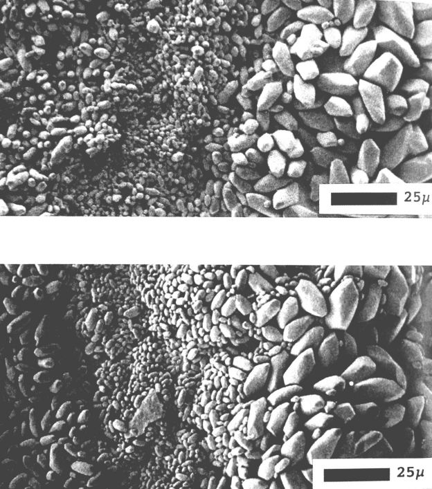
We observed no differences in the shape of the utricular otoconia and of the saccular otoconia between the 3 HG groups and the 2 matching CON groups. Furthermore, large, medium-sized and small otoconia were observed in the 3 HG groups and in the matching CON groups. We did not observe differences in density for each otoconial class (Fig. 1).
Fig. 1. Section of the utricular otoconial layer containing areas with large, medium-sized or small otoconia: a) patch of HG hamster (5 months in 2.5 G and 4 months in 1 G); b) patch of control hamster (9 months normal gravity).
Calcium content of the otoconia
The calcium content of small and large otoconia was the same, we observed no difference in the heights of the peaks. The calcium content of the HG groups did not differ from that in the matching CON groups, peak heights were the same for all groups. The otoconial calcium content of 1-month old hamsters was similar to that of 9-months old animals (Fig. 2).
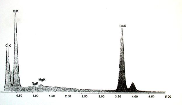
Fig. 2. Element analysis of the utricular otoconia: blackline = 1-month old HG hamsters; gray area = CON hamster (1 month normal gravity). Otoconial distribution on the utricular patch.
The otoconial distribution of the 3 HG groups
were compared to age-matched groups of controls. Group 1 (1-month old): the
otoconia distribution on the patches indicated that the area of the small otoconia
increased (t(15)=3.71, p=0.002) compared to the corresponding areas of the 1-month
old CON hamsters (Fig. 3).
Group 2 (9 months old: 5 months HG, 4 months 1 G): the otoconia distribution
on the pat- ches indicated that the area of the large
otoconia decreased (t(33)=5.07, p<0.001) and the area of the medium-sized
otoconia increased (t(33)=4.76, p<0.001) compared to the corresponding areas
of the 9-months old CON hamsters.
Group 3 (9 months old: 1 month HG, 8 months 1 G): the otoconia distribution
on the patches indicated that the area of the large otoconia decreased (t(28)=3.25,
p=0.003) and the area of the medium-sized otoconia increased (t(28)=2.89, p=0.007)
compared to the corresponding areas of the 9-months old CON hamsters (Fig. 4).

Fig. 3. Otoconial distribution of large, medium-sized and small otoconia on the utricular patch in hamsters (1 month old). HG = hypergravity hamster. Shown are means and standard deviations.
Age-related otoconial distribution and duration of HG exposure
No significant differences in otoconial distribution were found between the 1-month old and the 9-month old CON hamsters. The 1-month old HG hamsters (group 1) had a larger area with large otoconia (t(21)=3.70, p=0.001) and smaller areas with medium-sized (t(21)=4.75, p<0.001) and small otoconia ((t(21)=3.31, p=0.004) than group 2. Furthermore, the 1-month old HG hamsters had a smaller area with medium-sized (t(16)=2.60, p=0.02) and small otoconia (t(16)= 2.53 , p=0.025) than group 3.
Concerning duration of HG exposure after birth, we found no significant differences between 9-month old hamsters raised for 5 month under HG and 4 months under 1 G (group 2) and 9-month old hamsters raised for 1 month in HG and 8 months under 1 G (group 3).
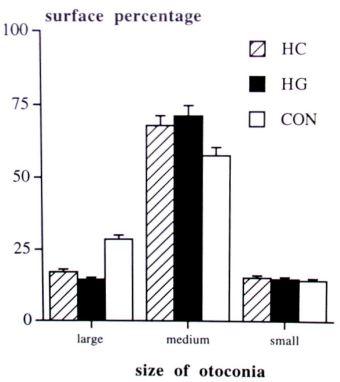
Fig. 4. Otoconial distribution of large, medium-sized and small otoconia on the utricular patch in adult hamsters (9 months old). Group 2 = HG hamsters (5 months HG and 4 months 1 G), Group 3 = HC hamsters ( 1 month HG and 8 months 1 G) CON = control hamster (9 months 1 G). Shown are means and standard deviations.
Discussion
Since the development of the peripheral
vestibular organs occurs in the embryonic period, it is suggested that hypergravity
affects the otoconial structure in such a way that the vestibular sensory input
becomes functionally appropriate for the increased G-load. Alterations have
been reported in the size or distribution of the otoconia (Lim et al. 1974;
Krasnov, 1991; Pedrozo and Wiederhold, 1994; Hara et al. 1995).
A delay in the formation of otoconia is found in chick embryos developed under
HG, which is probably caused by either an inhibition of Ca2+ deposit
or a change of otoconial precursors (Hara et al. 1995). The calcium incorporation
occurs mainly during the formation of the otoconia (Salamat et al. 1980). A
loss of calcium content is found during otoconial degeneration which is caused
by ageing or by labyrinthine diseases, resulting in behavioural disturbances
Ross et al. 1976; Johnsson et al. 1982; Campos et al. 1990). There is still
difference of opinion about a constant calcium exchange between the otoconia
and the otoconial membrane in mature otoconia. Ross (1979) found in rats a Ca2+
uptake by otoconia while Kawamata and Igarashi (1995) found no turnover of otoconial
calcium. If calcium exchange exists in mature otoconia, hypergravity could have
an effect on the otoconial calcium content. We found no difference in calcium
content between the hamsters of the HG groups and the matching CON groups; the
otoconial calcium content was identical in weaned and adult hamsters. This indicates
that an altered deposition or release of Ca2+, does not occur in
otoconia of animals developed under HG. Furthermore, we earlier observed no
difference in otoconial calcium content in hamsters exposed to HG for 6 months
after weaning (Sondag et al. 1995). Therefore, we conclude that exposure to
a new gravity condition does not alter the otoconial calcium exchange, neither
in hamsters born and conceived in HG and nor in hamsters exposed to HG after
weaning.
As far as the otoconial size is concerned, Krasnov (1991) found that the large
otoconia in the utricular peripheral zone were substituted by medium-sized ones
in 60-days old rats, gestated and raised under 2 G. We did not observe a lack
of large otoconia in our animals, they were present in both HG animals and controls.
However, we found changes in the relative area occupied by the large, medium-sized
and small otoconia on the utricular maculae; 1-month old HG hamsters had an
increased area with small otoconia and showed a trend towards a smaller area
containing large otoconia (not significant). The 9-month old HG hamsters had
a decreased area with large otoconia and an increased area with medium-sized
ones. We observed no differences in the density for each otoconial class area.
Assuming that the otoconial mass for each otoconial size class is different
from the other ones, we conclude that hypergravity causes a change in the otoconial
mass distribution on the utricular patch. This confirms the conclusion of Krasnov
(1991) that a change at the otoconial level of the utricular patch is one of
the mechanisms underlying the alteration in the sensory transduction of the
otolith organ upon changes in the gravity level.
As far as age-related differences are concerned, the 1-month old HG hamsters
(group 1) had a larger area with large otoconia than group 2 and a smaller area
with medium-sized and small otoconia than group 2 and 3, whereas no differences
were found between the 1-month old and 9-month old CON hamsters. These results
and the differences between HG hamsters and age-matched CON hamsters suggest
that the formation of the area with large otoconia is complete in the 1-month
old HG hamsters, while the "medium-sized area" is still developing.
Therefore, parts of the utricular otoconial layer in HG hamsters have a delayed
growth and are not yet fully matured one month after birth. This delayed growth
is in accordance with the results of Hara et al (1995) who found a delayed formation
of utricular otoconia in chick embryos developed under hypergravity. This delay
in otoconial formation might explain the differences we have found in the relative
size of the areas.
The duration of HG exposure after birth did not have an effect on the relative
areas occupied by the large, medium-sized and small otoconia; we found no differences
between HG hamsters who were raised under HG for 5 months (group 2) and animals
raised under HG for 1 month (group 3). Furthermore, changes in the relative
areas only appeared during pre- and post-natal development under HG conditions;
hamsters conceived and born under 1 G and subjected to HG for 6 months after
weaning showed no differences with controls (Sondag et al. 1995).
Based on our findings, we conclude that exposure to HG during the formation
of the otoconia does not alter the Ca2+ content, size or shape of
the otoconia. However, the relative areas occupied by large, medium-sized and
small otoconia on the utricular macula changes and the growth of some areas
seems to be delayed because of a structural adaptation to hypergravity. Differences
in otoconial distribution still exists after 8 months of normal gravity which
suggest that this adaptation is irreversible.
Acknowledgements
The authors gratefully acknowledge the Netherlands Organization for Scientific Research (NWO) for funding this project. This research was conducted while HNPM Sondag was supported by a grant of the Foundation for Behavioural and Educational Sciences (SGW) of this organization (575-62-049), awarded to Prof. Dr. WJ Oosterveld. We thank mr. FAW Peek (medical student, University of Amsterdam) for his assistance with the histological research. The authors also wish to thank mrs. L Kok-Noorman (scientific editor, Academic Medical Center) for her comments and manuscript review.