 Fig
1: Click to enlarge
Fig
1: Click to enlargeAbstracts NL-Symposium Symposium, Friday 10th December 1999.
Free University Amsterdam, ĹTransitoriumĺ building
room KB-74, van der Boechorststraat 1, Amsterdam.
Click
here for travel direction to the Free University.
MICROGRAVITY RESEARCH IN EUROPE, PAST PERFORMANCE AND NEW CHALLENGES.
Prof.Dr. E.H. Burger
Dept. Oral Biology, Sect. Cell Biology, ACTA-VU, van der Boechorststraat 7, 1081 BT Amsterdam, The Netherlands. Tel: +31 (0)20 4448661, E-mail: eh.burger.ocb.acta@med.vu.nl.
In her presentation Prof. Burger mainly addressed three points:
Number of papers by a European author of co-author (dark blue line) in relation to the a measure for scientific quality of the journal in which the papers are published (the impact factor, IF) (light blue line).
In the above graph, an extrapolation of the number of publications for the years 1999-2000 has been plotted. The impact factor in 1999-2000 corresponds to the current average impact factor for the year 1999.
MICROTUBULE SELF-ORGANISATION DEPENDS UPON GRAVITY.
J. Tabony, N. Pochon, C. Papaseit
Laboratoire R.M.B.M, Département de Biologie Moléculaire et Structurale, D.S.V, C.E.A. Grenoble,17 rue des Martyrs, 38054 Grenoble Cedex 9, France. jtabony@cea.fr
The molecular processes by which gravity is transduced into biological systems
are poorly, if at all, understood. Under equilibrium conditions, chemical and
biochemical structures do not depend upon gravity. It has been proposed that
biological systems might show a gravity dependence by way of the bifurcation
and self-organising properties of certain types of non-linear chemical reactions
that are far-from-equilibrium. We have found that in-vitro preparations of microtubules,
an important element of the cellular cytoskeleton, show this type of behaviour.
On earth, the solutions show macroscopic self-ordering, and the morphology of
the structures that form depend upon the orientation of the sample with respect
to gravity at a critical moment prior to the progressive appearance of the self-organised
state. An experiment carried out in a sounding rocket, showed that as predicted
by theories of this type, no self-organisation occurs when the microtubules
are assembled under microgravity conditions. The same preparations formed on
a 1g on-board centrifuge showed the morphologies that form on earth. This is
an experimental demonstration of how a very simple biochemical system, containing
only two molecules, can be gravity sensitive. At a molecular level this behaviour
results from an interaction of gravity with macroscopic concentration and density
fluctuations that arise from the processes of microtubule contraction and elongation.
In addition, the self-organising process is capable of transporting particles,
such as colloidal beads, chromosomes, nuclei, etc., along a given direction
at rates of several microns per minute. Particle movement results from collective
processes of microtubule assembly and disassembly involved in the self-organising
process. No particle motion occurs when microtubules are assembled under conditions
such that self-organisation does not occur. As microtubule self-organisation
does not occur under microgravity conditions, then microgravity should also
inhibit particle movement arising from microtubule self-organisation.
Click here for actual presentation.
EFFECTS OF GRAVITY ON MOLECULAR SELF-ASSEMBLY
Köhler Gottfried(1,2), Rünzler Dominik(1,2), Mayer Bernd(2), Sára Margit(3) and Rasmussen Steen(4)
1 Institute for Space Biophysics, Austrian Society for Aerospace Medicine-ASM, Lustkandelgasse 52/3, A-1090 Vienna, Austria; A-1090 Vienna , Austria, Tel.: 0043 1 3155777
Fax: 0043 1 31557778, E-mail: headoffice@asm.at; 2 Institute for Theoretical Chemistry and Structural Biology, University of Vienna, A-1090 Vienna; 3 Center for Untrastructure Research, Agricultural University, and Ludwig-Boltzmann-Institute for Molecular Nanotechnology, A-1180 Vienna; 4 EES-5 MS D407 and T-CNLS MS B258, Los Alamos National Lanoratory, Los Alamos, New Mexico, USA
Key words: organic materials, microgravity, molecular nanotechnology, protein crystallization, oligomerization
Experimental and computer modeling investigations are performed
with the aim to understand the influence of gravity on molecular self-assembly
of organic and bioorganic materials. Important issues are to improve the quality
and homogeneity of protein crystals and electronic and photonic materials. The
understanding of particle nucleation, growth, aggregation and dispersion is
an important prerequisite for the production of nanoscale partices for nanotechnological
applications.
Molecular self-assembly covers in general the emergence of supramolecular structures
with defined functionality on a nano- or microscopic scale from pure physico-chemical
properties of their constituents. Protein folding, i.e. the process leading
from the primary sequence of amino acids to the native functional state of the
protein, follows the same concepts than molecular self-assembly. It is a fascinating
task to apply the same organizational concept for the developement of novel
materials. Two or three dimensional ordering of the molecular constituents is
an important feature in self-assembly of such supramolecular structures. Such
materials comprise liquid crystals, organogels or hydrogels, etc. but they have
important objectives for the developement of nanostructured materials, i.e.
of materials with function on a nanometer scale. Applications for miniaturization,
for optical or electronic data storage or processing are the main issues for
the applications of such materials. Sedimentation is an important effect often
limiting the growing of assemblates due to gravity. In a macromolecular solution
the initially homogeniously distributed molecules or aggregates are redistributed
along the gravity gradient depending on the density of the particles. Gravity
is generally a source of convection and deteriorates self-assembly and crystallization.
Conventional models applied to describe structure and dynamics of large molecular
systems use an atomistic description within a force field. Optimization of molecular
structure is then performed via local minimization methods, as e.g. gradient
minimizers and molecular. However, these methods are severely restricted concerning
system size as well as the tracktable time interval which can be computed in
molecular dynamics. Therefore, for the studying self - assembly of large molecular
ensembles over a long time scale, which is necessary for the systems described
above, requires a different type of description and one successful example is
the Lattice Molecular Automaton (LMA) which will be discussed in detail.
The threading of macrocycles on chain polymers or the formation of cylindrical
bundles of appropriate molecules hold together by macrocycles are considered
as excellent model systems for the formation of nanotubes or nanowire as naotechnological
devices. The formation of such supramolecular structures has been demonstrated
using cyclodextrins, which are cyclic oligosaccharides, as macrocycles which
are threaded on block copolymers form rigid rods with highly polarizable molecules.
Another example it the assembly of bacterial surface layer proteins which form
uniform layers, structured on a nanometer scale. The influence of gravity on
such self-assembly processes is shown and the importance of physico-chemical
studies under microgravity is discussed.
SIMULATED MICROGRAVITY ON THE RANDOM POSITIONING MACHINE AND THE RESPONSES OF ISOLATED FETAL MOUSE LONG BONES
J.Paul Veldhuijzen, Jolanda de Blieck-Hogervorst
ACTA, dept. Oral Biology, van der Boechorststraat 7, 1081 BT Amsterdam. The Nehterlands. Tel: 020-4448664, Fax: 020-4448683, E-mail: J.V.Veldhuijzen.ocb.acta@med.vu.nl.
Microgravity has severe effects on the musculo-skeletal system. Both astronauts and animal models have shown that bone mass is rapidly reduced during space flight. Under in vitro conditions it has also been shown that bone cells respond to microgravity. We have demonstrated that in fetal mouse long bones 4 days of microgravity significantly reduced the mineralization of the hypertrophic cartilage, whereas growth was not affected. Recently ground based system such as the Random Positioning Machine (RPM) has been developed which provide simulated microgravity. In this type of a machine, which is basically a 3D-clinistat, it has been reported that in plant models and animal cells (lymphocytes) real microgravity responses could be duplicated, suggesting that the RPM may be a good tool in the ground based microgravity research. In a series of experiments we have tried to reproduce the effects found in real microgravity on the growth and mineralization in isolated fetal mouse long bones in order to show that the RPM indeed generates simulated microgravity. In these experiments we have used 17-day-old fetal mouse long bones, which were cultured for 4 days in the same hardware as used in the actual flight experiments. The long bones were cultured in polyethylene culture bags (Berlingots) in 0.7 ml of medium. Culturing long bones free floating the medium of the Berlingots for 4 or 5 days on the RPMN did not change the growth and mineralization. This may be due to the movement of the long bones in the Berlingots caused by the RPM rotation. These unwanted movements could have interfered with the necessary random movement of the RPM to provide simulated microgravity. To avoid these several techniques were used to immobilize the long bones. Alginate-gels were applied but presumably due to a high concentration of Ca2+ ions, necessary in the preparation of the gels, mineralization was very strongly stimulated, masking any possible effects of the RPM on mineralization. Growth was not or marginally affected. We also tried a specific tissue glue (matrigell), however application on the isolated long bones was very difficult, resulting in a very bad growth and even death of the long bones. We finally decided to embed the long bones in a thin layer of agarose, which had minor negative effect on growth and mineralization. Culturing long bones immobilized in agarose on the RPM however gave very fluctuating results. Growth was not affected, however the effects on mineralization were very variable. The fluctuating results could be caused by the difficulty to avoid air bubbles in the system. Air bubbles, especially on the RPM, will cause better mixing of the medium which is not found in the controls. However a decreased mineralization seemed not to be correlated with the absence of air bubbles. It is concluded that we were not able to reproduce our flight results with fetal mouse long bones on the RPM. This may be partly due to unforeseen difficulties in adapting our proven culture system for flight experiments for use on the RPM. Therefore the result of this pilot project is undeceived in demonstrating the usefulness of the RPM in providing simulated microgravity for cultured fetal mouse long bones. (supported by SRON through grant MG-047)
EFFECT OF RED AND BLUE LIGHT ON THE TIMING OF CELL DIVISION IN THE UNICELLULAR GREEN ALGA CHLAMYDOMONAS REINHARDTII
Oldenhof Harriëtte, v.d. Ende Herman
Institute for Molecular Cell Biology, section Plant Physiology, Biocentrum Amsterdam, University of Amsterdam, Kruislaan 318, 1098 SM Amsterdam, The Netherlands. Tel: 020-5257846, e-mail: oldenhof@bio.uva.nl
key words:cell cycle, Chlamydomonas
Chlamydomonas reinhardtii is a multiple fission alga, which implies
that it produces a multiple number of two daughter cells per mother cell after
one G1 phase. During this G1 phase critical cell sizes are passed which are
sufficient for the completion of the cell cycle.
We are interested in the regulatory mechanism underlying this process, so that
we can explain differences in proliferation at different conditions. Under ordinary
white light conditions cell divisions occur after a constant time after the
onset of the light period. Cultures grown in the presence of only blue light
show a later timing of cell division when compared with cultures grown in the
presence of only red light. We discuss the involvement of light perception in
the uncoupling of the initiation of cell division from cell growth and resulting
cell sizes.
BIOACTIVE GLASS STIMULATION OF EMBRYONIC BONE MINERALIZATION IN SIMULATED MICRO-GRAVITY CONDITIONS
Maroothynaden J., Hench L.L.
Centre for Tissue Engineering, Department of Materials, Imperial College of Science, Technology and Medicine, Prince Consort Road, London SW7 2BP. Tel. +44(0)171-594-6813, Fax. +44(0)1715946809, Email: jm2@ic.ac.uk
Keywords: Bioactive glass, femora, long-bone, simulated micro-gravity, mineralisation, Random Position Machine.
Load bearing bones demineralise at much faster rates in space
than on the Earth. Even though this phenomenon has been quantified, the fundamental
biological mechanisms remain unresolved. Also, the chemical effects responsible
for enhanced bone formation observed in-vitro and in-vivo at 1-gravity
with bioactive glasses may be sufficient to prevent demineralisation that is
occurring in micro-gravity or as a consequence of age or surgery.
Embryonic mice femora were cultured under two conditions - a supplemented alpha-MEM
control medium and in a bioactive glass extract medium - in a Ĺsimulated micro-gravityĺ
environment provided by the Random Position Machine (Dutch Experiment Support
Centre, Holland) for 4-days. A simultaneous 1-gravity control was performed.
Femora cultured in the Ĺsimulated micro-gravityĺ environment without bioactive
glass stimulation followed similar reported histomorphometric trends as long
bones cultured in-vitro during space-flight. Decreased mineralisation
was observed along with a change in embryonic growth-plate kinetics. For the
'simulated micro-gravity' cultures where the femora were stimulated chemically
to a bioactive glass extract solution, the negative effects of Ĺsimulated micro-gravityĺ
were retarded. In-fact there was no statistical change in mineralisation rates
between the Ĺsimulated micro-gravityĺ bioactive glass extract exposed cultures
and the control 1-g cultures were followed. The positive effect of the bioactive
glass extract on mineralisation was also seen in the corresponding 1-g cultures
but at a more accelerated rate.
We have developed an easy to use and repeatable in-vitro embryonic mouse
long-bone model that makes it possible to examine the coupling of biochemistry
and biomechanics inherent to bone health thereby enhancing understanding of
the mechanisms involved in demineralisation. We have also demonstrated that
bioactive glasses, in extract form, may retard and even reverse Ĺsimulated micro-gravityĺ
induced demineralisation.
Click here for actual presentation.
EFFECT OF SIMULATED MICROGRAVITY ON DENDRITIC CELL MIGRATION AND MATURATION
Vermaelen Karim, Romain A. Pauwels
Dept. of Respiratory Diseases 7K12ie Ghent University Hospital De Pintelaan 185 B-9000 Ghent Belgium tel: 32 9 2402605 fax: 32 9 2402625, E-mail: Karim.Vermaelen@rug.ac.be
Keywords: microgravity, dendritic cell, DTH (delayed type hypersensitivity), chemotaxis
Space travel has been shown to affect the immune system. Although
serious clinical manifestations have been absent so far, several observations
have pinpointed disturbing anomalies.
Many studies have uncovered an impairment of T lymphocyte activation after exposure
to real or simulated microgravity, both in vivo and in vitro. Moreover, a depressed
cutaneous delayed type hypersensitivity reaction (DTH) has been observed after
space flight. There are reasons to believe that environment-specific, stress
hormone-independent factors are contributing to immune depression in space.
Like any other antigen-specific immune response, the DTH does not only rely
on intact T-cell function. Indeed it is now widely recognized that, on a more
upstream level, antigen-presenting dendritic cells (DCs) have to perform adequately.
These cells are strategically positioned in the skin or at other sites of high
antigen exposure. The crucial sentinel function of DCs depends on a perfectly
coordinated migration of these cells from the antigen-exposed surface to the
T-cell areas of draining lymph nodes. This migration relies on adequate changes
in cell shape and adhesive properties in response to very specific chemotactic
signals. Microgravity has been shown to disturb cytoskeletal organization (Hashemi
et al., manuscript in preparation). Therefore, it is possible that Ág-induced
cytoskeletal dysfunctions could hamper correct antigen delivery by migrating
DC and thus depress the DTH. The latter phenomenon is only a warning flag of
a disturbed cellular immune response as a whole. This may well be crucial in
the context of future long duration space stays in which high exposure to radiation
increases the burden on our immunological tumor surveillance system. DCs play
a central role here as well: they are the only cells known to effectively prime
antitumor cytotoxic T lymphocytes. Therefore in response to concerns about crew
health in space, it would be interesting to observe whether the two key dendritic
cell properties, i.e. maturation and directed migration, are disturbed in microgravity.
For the experiments proposed, large amounts of differentiated dendritic cells
will be obtained from mouse bone marrow cell cultures in the presence of Flt3-L,
GM-CSF and IL-4. Following this culture period, experiments will be performed
in a RPM vs 1-g control. For obvious reasons, incubations will not proceed in
a liquid phase, but rather cells will be entrapped within a collagen gel matrix
prior to exposure to simulated Á-g. This will also eliminate differences in
cell sedimentation rates (influencing cell-cell interactions) between the two
groups as a potential interfering factor.
A first set of experiments will involve a chemotaxis assay using a potent DC
chemoattractant (e.g. fMLP) and DCs seeded into one of two filter-separated
collagen gels (a checkerboard analysis will distinguish genuine chemotaxis from
chemokinesis). In a second set of experiments, DCs will be incubated overnight
in a collagen gel, in the presence or absence of an additional maturational
stimulus (e.g. LPS). After enzymatic gel dissolution, maturation will be assessed
by flow cytometry using monoclonal antibodies against MHCII and a selected battery
of costimulatory molecules.
Click here for actual presentation.
ANIMAL
HYPERGRAVITY RESEARCH session.
Chairperson: Jack J.W.A. van Loon
In this
session data are presented that emerged out of the 'tissue sharing' program
from the animal centrifuge located at the Academic Medical Center (AMC) in Amsterdam.
In this program various research groups use the same animals to investigate
changes in e.g. vestibular system, bone, muscle, collagen, heart etc., due to
2.5Îg hypergravity. See picture
for overview.
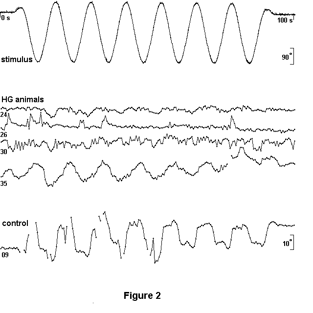
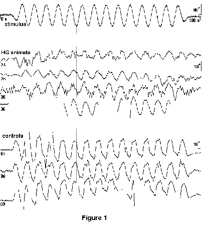
THE VESTIBULO-OCULOMOTOR REFLEX OF HYPERGRAVITY RATS
Wubbels, René J., de Jong, Herman A. A.
Vestibular Department ENT, Academic Medical Center, University of Amsterdam, PO Box 22660, 1100 DD Amsterdam, The Netherlands. e-mail: r.j.wubbels@amc.uva.nl
Keywords: rat, hypergravity, centrifuge, vestibular system, vestibulo-oculomotor reflex
Long-Evans rats were bred in our laboratory. One group lived
under normal gravity conditions. Another group were conceived and born, and
thereafter lived, at hypergravity (HG) in a centrifuge. Rotation at 34.3 cycles/minute,
resulting in a gravity level of 2.5 g, was alternated from clockwise to anti-clockwise
(or vice versa) every day. The vestibulo-oculomotor reflex (VOR), which compensates
for motion of head and body in order to stabilize the visual image on the retina,
was measured for both groups. The animal was immobilized in a light-tight box
on a (horizontal) platform with its labyrinths positioned approximately at the
platformĺs axis of rotation. The position of the (left) eyeĺs pupil (angular
resolution: 0.2░ ) was monitored with a camera (IR illumination) and recorded
on VCR (temporal resolution: 20 ms), together with the applied stimulus.
In one type of experiment, the rats were sinusoidally oscillated (T=6s; ampl.=90░
). The response of 4 HG animals and 3 controls is shown in Fig. 1. The upper
trace represents the stimulus. Interruptions of the response trace indicate
a closed eye. Numbers at the left-hand side of the response traces represent
individual animals. Scale bars: 90░ for stimulus, 10░ for responses. Temporal
resolution: 500 ms. Fig. 2 shows the result of a similar type of experiment
(T=12s; ampl.=180░ ) with 4 HG animals and one control.
The general conclusion, from experiments in which we applied an angular acceleration,
is that the amplitude of the VOR of HG animals is smaller than that of control
animals. This also holds for the nystagmus (a component of the total VOR). Superimposed
on compensating eye movements like those shown in Figs. 1 and 2, a nystagmus
can be observed in the response of some of the control animals; in the response
of HG rats we saw no nystagmus. Finally, there is a phase difference between
the responses of HG and control animals (see Fig. 1). The distortion of the
sinusoidal compensatory eye movement is caused by physical constraints which
limit the eyeĺs rotation in its socket (constraints which this sensory-motor
system attempts to overcome by the nystagmus component of the VOR).
The origin of the smaller VOR amplitude of HG rats (measured under 1 g conditions)
remains a matter of speculation. Freely moving animals in a rotating system
like our centrifuge are subject to Coriolis forces which depend on the motion
vectorĺs magnitude and on its angle with the plane of rotation. It is possible
that under these conditions, the vestibularly detected acceleration is a less
reliable source for anticipating the accompanying movement of the visual field
across the retina.
THE CONSEQUENCES OF LONG TERM EXPOSURE TO HYPERGRAVITY FOR BONE MASS IN RATS.
Bravenboer Nathalie, Huib van Essen, Annechien Tromp, Adrien Krui.
University Hospital of the Free University Amsterdam, Dept. Endocrinology, De Boelelaan 1117, 1081 HV Amsterdam, Tel: 020-4442641, Fax 020-4444313. E-mail: n.bravenboer@azvu.nl .
The importance of mechanical
loading for maintaining skeletal integrity is well established. Our goal is
to investigate the biological response to mechanical loading which results in
a higher bonemass. Using the rat with backpack-model we showed that a short
duration of exercise with a high additional load stimulates bone mass, as determined
by dual X-ray absorptiometry (DXA). Biomechanical competence was also stimulated
by additional weight-bearing (1).
Additional weight-bearing during exercise also resulted in a modulation of the
bone concentrations of the growth factors IGF-I and TGF-▀. However serum IGF-I
and GH remained constant. In the model of the rat living under hypergravity
conditions, we wanted to investigate the effect on bonemass by DXA (VU-Amsterdam)and
by histomorphometry, (Dr Zerath and Dr. Holy, France). In addition to these
tests bones were also tested for structure and biocompetence using microCT,
which was performed in Nijmegen (see Abstract Tanck et al.) and by biomechanical
bending tests, which were performed by Dr. Mosekilde, Aarhus, Denmark. Furthermore,
we measured the concentration of IGF-I and IGFBP3 in tibiae. DXA scans of extracted
tibiae did not show any significant differences between hypergravity and control.
At the moment we are performing total body DXA-scans of hypergravity and control
animals. Preliminary data of 10 rats show that BMC and BMD as well as bodyweight
and lean body mass do not differ between the two groups in female rats. However
fatmass measured in grams (p<0.01) and as percentage of bodyweight (p<0.01)
is significantly lower in the hypergravity female rats compared to the control
female rats.
In conclusion so far: Bonemass and bonecomposition (IGF-I and TGF-▀) are unchanged
under hypergravity conditions. Hypergravity reduces fatmass but not total bodyweight
in female rats.
(1) Van der Wiel et al., Bone 16: 73-80 (1995).
CHRONIC HYPERGRAVITY INCREASES THE CONTENT OF HEAT SHOCK PROTEIN HSP70 IN SKELETAL MUSCLES OF RATS
Van Nieuwenhoven Frans A., Heijnen Viviane V.T., De Jong Herman A.A. and Snoeckx Luc H.E.H.
Department of Physiology, Cardiovascular Research Institute Maastricht (CARIM), Maastricht University, P.O. Box 616, 6200 MD Maastricht, the Netherlands. Phone +31-43-3881212, Fax +31-43-3884166, E-mail F.vanNieuwenhoven@fys.unimaas.nl, and Vestibular Department, Academic Medical Center (AMC), Amsterdam, the Netherlands.
Key words: HSP70, hypergravity, skeletal muscle, rat
Introduction: Environmental stresses such as heat, toxins or
hypoxia, cause tissues to express a special set of genes leading to increased
levels of so-called heat shock proteins (HSPs). This is a universal process,
which can transiently protect tissues against a subsequent stressful event.
One of the strongest induced HSPs in mammalian tissues is HSP70, which is a
member of the 70-kDa HSP-family. Since it is essentially unknown whether chronic
hypergravity (HG) exerts stress upon tissues, the expression of HSP70 was studied
in several tissues of adult HG-rats.
Methods: Three male Long Evans rats were conceived, born and raised in a large
radius animal centrifuge under 2.5 times g force, and three age-matched control
rats were kept under 1 time g force. At the age of 7 months the rats were sacrificed
and the following tissues harvested: heart, brain, lung, liver, kidney, testes,
soleus muscle and gastrocnemius muscle. Tissues were homogenized and total protein
content was determined. Thereafter homogenates were boiled in SDS-PAGE sample
buffer to denature all proteins. Following SDS-PAGE and Western blotting, HSP70
was identified using a specific polyclonal antibody. After chemiluminescent
detection, the signals from the Western blot analyses were quantified using
a CCD camera (Fluor-S Imager, Biorad) and compared to known standards.
Results: A signal for HSP70 could be detected in all tissues of control and
HG rats examined, thus showing that although this protein is described to be
strongly induced upon a number of different stress situations, it is also present
in the non-stressed situation. Soleus and gastrocnemius muscles showed an increased
HSP70 content in the HG rats compared to the control rats, while all other tissues
revealed no major differences.
Conclusion: Chronic hypergravity leads to increased HSP70 levels in soleus and
gastrocnemius muscles in rats, while in other tissues normal expression levels
were observed. The function of the increased amount of HSP70 in skeletal muscle,
however, remains elusive.
The authors wish to thank the Dutch Experiment Support Center (DESC) for facilitating
this research project
STUDIES ON RAT HEARTS SUBMITTED TO PROLONGED HYPERGRAVITY
M. de Jong, J.F. Jongkind, H.A.A. de Jong*, J.J.W.A. van Loon**, J.W. de Jong.
Thoraxcenter, Erasmus University Rotterdam, NL; *Vestibular Department ENT, Academic Medical Center, University of Amsterdam, NL; **Dutch Experimental Support Center, ACTA-Vrije Universiteit Amsterdam, NL. Correspondence: E-mail: j.w.dejong@tch.fgg.eur.nl
Keywords: centrifuge, heart, hypergravity, rat
Background. Microgravity induces substantial loss of skeletal muscle
mass. Few data indicate that atrophy occurs in heart muscle during weightlessness.
Substantiation and characterization of these findings are hampered by limited
experimental possibilities in space and inadequate simulation microgravity models
on Earth.
Hypothesis. Assuming that microgravity and hypergravity affect the heart
in an inverse way, we hypothesized that prolonged hypergravity induces hypertrophy
in cardiac muscle.
Experimental. Rats of either sex were raised in a centrifuge,
i.e., they were subjected to hypergravity (2.5 x g) for up to 16 months. A control
group of rats was treated similarly outside the centrifuge. The animals were
killed by decapitation. Their hearts were immediately removed, rinsed several
times in cold phosphate-buffered salt solution, weighed, and fixed by immersion
in cold phosphate-buffered 4% paraformaldehyde. Slides underwent light-microscopic
examination in a blinded fashion.
Preliminary results. In the groups of animals studied thus far, we were
unable to observe differences in the ratio heart weight/body weight. However,
the hearts obtained from the hypergravity group showed morphological signs of
hypertrophy, in contrast to the control hearts.
Conclusion. Further centrifugation studies and detailed morphometric
analyses are needed to confirm our preliminary finding that hypergravity induces
cardiac hypertrophy.
TRABECULAR BONE ARCHITECTURE IN THE RAT, NORMALLY AND AFTER LONG-TERM EXPOSURE TO 2.5 HYPERGRAVITY.
Tanck Esther, Van Lenthe G Harry, Huiskes Rik.
Orthopaedic Research Lab University of Nijmegen, P.O. Box 9101, 6500 HB Nijmegen, The Netherlands Tel: 024-3617536, Fax: 024-3540555, E-mail: esther@orth0044.azn.nlKey words: bone architecture, micro-CT, hypergravity
Exposure to hypergravity has shown to decrease body weight, femur mass and
femur size compared to controls2. The femur size relatively to body
weight, and the wall thickness of the shaft may increase however, resulting
in increased bone strength1,3,4. On the other hand, overloading may
immobilize the animal, resulting in decreased bone strength5. In
this pilot-study, we investigated the following question. What is the influence
of long-term exposure of 2.5 hypergravity to bone architecture in male rats?
With micro-CT, the 3D bone architecture was determined. Morphological parameters
like bone volume fraction (BV/TV), trabecular thickness (Tb.Th), ratio of bone
surface to bone volume (BS/BV), and degree of anisotropy, which is a measure
for trabecular orientation, were measured. Information about the mineral content
cannot be obtained.
Three-week old rats were exposed to 2.5 hypergravity for 38 weeks (HG-group-1;
N=2). Control rats were raised under normal conditions (N=2). In addition, rats
were procreated, born and raised under 2.5 hypergravity conditions for 33 weeks
(HG-group-2; N=3). The proximal femur of each rat was scanned in a micro-CT
with a spatial resolution of 28 micrometer. A 2x2x2mm3 volume of
interest of the trabecular region in the femoral head was segmented and evaluated
for the various morphological parameters.
BV/TV was similar for all groups (Table 1). For HG-1, but not for HG-2, trabecular
thickness was decreased by 6% and BS/BV was increased by 5% compared to controls.
Compared to controls, total body weight was decreased by 16% for HG-1 and 29%
for HG-2.
|
|
controls |
HG-1 |
HG-2 |
|
BV/TV |
0.58 ▒ 0.01 |
0.60 ▒ 0.01 |
0.62 ▒ 0.02 |
|
Tb.Th (micrometer) |
125 ▒ 0.7 |
118 ▒ 1.4 * |
125 ▒ 1.5 |
|
BS/BV (1/mm) |
16.1 ▒ 0.14 |
16.9 ▒ 0.14 * |
15.9 ▒ 0.21 |
|
degree of anisotropy |
1.32 ▒ 0.007 * |
1.28 ▒ 0.007 |
1.26 ▒ 0.012 |
|
total body weight (g) |
576 ▒ 2 * |
482 ▒ 21 * |
410 ▒ 10 * |
Table 1: Morphological parameters for the three experimental groups (mean ▒ SD). In addition, total body weight is included. * p<0.05 compared to the other groups.
These preliminary results indicate that the trabecular architecture
in the femoral head hardly changed when exposed to 2.5 hypergravity. It should
be noted that the HG-2 group was not age-matched compared to the other groups;
additional controls are required. Furthermore, the femoral head contains a growth-plate,
which might affect the results.
Bone adapts to mechanical load. It was therefore unexpected that the BV/TV did
not increase under hypergravity conditions. The cortical thickness, femoral
mass, and femoral length were, however, not measured but could be different
compared to controls1,2,3, so that adaptation to hypergravity conditions
might be more at the global, cortical level than at the trabecular level. On
the other hand, it is possible that the activity of HG-rats was less compared
to controls. This would result in decreased dynamic stimulation of the bone
so that the unchanged BV/TV still may satisfy the mechanical demands of the
rats exposed to hypergravity.
References 1) Gordon et al., Bone, 303-12, 1989.; 2) Keil et al., Physiol, 553-4,
1979; 3) Kimura et al., J Biomech, 361-5, 1979; 4) Wunder et al., Aviat Space
Environ Med, 339-46, 1977; 5) Wunder, Aviat Space Environ Med, 1023-25, 1977.
HYPERGRAVITY AFFECTS LYSYL HYDROXYLATION AND PYRIDINOLINE CROSS-LINKING IN RAT CORTICAL BONE
Ruud A. Bank(1), Tom van de Broek (1), Herman A.A. de Jong (2), Jack J.W.A. van Loon (3) & Johan M. TeKoppele(1)
TNO Prevention and Health, Divison of Vascular and Connective Tissue Research, P.O. Box 2215, 2301 CE Leiden, The Netherlands, phone +31-71-5181503, email RA.Bank@tno.pg.nl (2) Vestibular Department ENT, Academic Medical Center, University of Amsterdam, PO Box 22660, 1100 DD Amsterdam, The Netherlands. e-mail: r.j.wubbels@amc.uva.nl (3) Dutch Experimental Support Center, ACTA-Vrije Universiteit, Dept. of Oral Biology, Van der Boechorststraat 7, 1081 BT Amsterdam, phone +31-20-448664, email DESC@wxs.nl
Bone tissue is subjected to a continual process of resorption and renewal; this remodelling is operative throughout life. Osteoblasts synthesize the organic matrix of bone, which consists mainly of collagen type I. Synthesis of collagen molecules and assembly of the collagen molecules into a network of fibrils (accomodating most of the mineral phase of bone) requires a large number of intra- and extracellular post-translational modifications. Key steps in the formation of a biomechanically strong collagen network are lysyl hydroxylation of the triple helix and cross-linking of the telopeptides. It has only recently been appreciated that the quality of the collagen network plays an important role in the pathogenesis of osteoporotic fractures. Unfortunately, in contrast to the mineral phase of bone, little attention has been paid so far to the structure of the collagen network, being the reason that we know little about the posttranslational chemistry of collagen even in normal bone. Our studies on rat bone revealed that large differences occur in the enzymatically mediated modifications of collagen (lysyl hydroxylation and cross-linking), not only between cortical and trabecular bone, but also within cortical bone derived from various regions of the same diaphysal shaft. Apart from that, age-related changes are seen, and it was found that lifetime caloric restriction is able to modulate the enzymatic modifications of collagen. Clearly, bone accomodate phenotypically different osteoblast populations. As yet, little is known about the factors that modulate this differentiation. Apart from soluble factors (such as hormones and cytokines), mechanical factors are likely to play a role. To investigate the latter, we have studied the properties of the collagen network in cortical bone of the proximal part of the femur and the tibia of rats subjected to chronic hypergravity (2.5 g; 10-16 months) and compared it with age-matched controls. In addition, the same was done with collagen derived from tendons of the tail. No significant differences were found in lysyl hydroxylation and cross-linking in tail tendon. In contrast, the collagen network in bone revealed significant differences in hypergravity rats. The proximal part of the femur showed, compared to controls, a lower level of lysyl hydroxylation of the triple helix as well as a lower amount of the cross-links hydroxylysylpyridinoline (HP) and lysylpyridinoline (LP) per collagen molecule; the HP to LP ratio did not change. The proximal part of the tibia showed, compared to controls, a lower level of lysyl hydroxylation of the triple helix, a lower amount of LP per collagen molecule and a higher HP to LP ratio. Evidently, rats that live under hypergravity conditions show an altered bone collagen network. As lysyl hydroxylation of the triple helix is an intracellular process, it can be concluded that osteoblasts in hypergravity rats are phenotypically different from that of control rats.
MICROGRAVITY AND BONE CELL MECHANOSENSITIVITY
Klein-Nulend J, Veldhuijzen JP, Van Loon JJWA, Burger EH
ACTA-Vrije Universiteit, Dept Oral Cell Biology, Van der Boechorststraat 7, 1081 BT Amsterdam, The Netherlands. tel.: +31(0)20-4448667, Fax: +31(0)20-4448683, e-mail: j.klein_nulend.ocb.acta@med.vu.nl
Key words: Microgravity, Bone cells, Mechanosensitivity, Fluid flow, Prostaglandins, Nitric Oxide
The capacity of bone tissue to alter its mass and structure in response to
mechanical demands has long been recognized but the cellular mechanisms involved
remained poorly understood. Bone not only develops as a structure designed specifically
for (future) mechanical tasks, but it can adapt during the life of an individual
toward more efficient mechanical performance. Mechanical adaptation of bone
is a cellular process and needs a biological system that senses the applied
mechanical loading. The loading information must then be communicated to the
effector cells that can make new bone or destroy old bone.
The in vivo operating cell stress derived from bone loading is likely
flow of interstitial fluid along the surface of osteocytes and lining cells.
The response of bone cells in culture to fluid flow includes prostaglandin synthesis
and expression of prostaglandin G/H synthase inducible cyclooxygenase (COX-2).
Cultured bone cells also rapidly produce nitric oxide (NO) in response to fluid
flow as a result of activation of endothelial nitric oxide synthase (ecNOS),
which enzyme also mediates the adaptive response of bone tissue to mechanical
loading. Disruption of the actin-cytoskeleton abolishes the response to stress,
suggesting that the cytoskeleton is involved in cellular mechanotransduction.
Microgravity, or better near weightlessness, has catabolic effects on the skeleton
of astronauts, as well as on mineral metabolism in bone organ cultures. This
might be explained simply as resulting from an exceptional form of disuse under
near weightlessness conditions. However, under near weightlessness conditions
the assembly of cytoskeletal elements may be altered since it has been shown
that the direction of the gravity vector determines microtubular pattern formation
in vivo. We recently found that the transduction of mechanical signals
in bone cells also involves the cytoskeleton and is related to PGE2
production. Therefore it is possible that the mechanosensitivity of bone cells
is altered under near weightlessness conditions, and that this abnormal mechanosensation
contributes to the disturbed bone metabolism observed in astronauts.
In a recently submitted project proposal in response to the International Life
Sciences Research Announcement for the Utilization of the International Space
Station (ESA-AO-98-LSRA), we wish to test this hypothesis experimentally using
an in vitro model. The specific aim of our research proposal is to test
whether near weightlessness decreases the sensitivity of human bone cells for
mechanical stress through a decrease in early signaling molecules that are involved
in the mechanical loading-induced osteogenic response. A human bone cell line
is cultured with or without gravity prior to and during mechanical loading.
Mechanical loading will be applied using our modified in vitro oscillating
fluid flow apparatus. To exclude that results are confounded by reduced mass
transport during near weightlessness, controls will be treated by low fluid
flow, representing baseline levels of mechanical stress. Cell culture conditions
and cell responses will be measured on line using, glucose / lactate sensors
and nitric oxide sensors, respectively. At the end of the experiment conditioned
medium will be tested for prostaglandin and nitric oxide production. Semi-quantitative
polymerase chain reactions will be performed to study COX and NOS mRNA expression.
This study will develop a cell culture module that is used to provide further
insight in the mechanism of mechanotransduction in bone.
EVOLUTION OF ORGANIC MATTER IN SPACE - INTERNATIONAL SPACE STATION ISS
Ehrenfreund Pascale, Ruiterkamp Richard.
Raymond and Beverly Sackler Laboratory for Astrophysics at Leiden Observatory, Niels Bohrweg 2
P.O. Box 9513, 2300 RA, Leiden, The Netherlands, tel: 0715275812, fax: 0715275819, e-mail: pascale@strw.leidenuniv.nl, richard.ruiterkamp@strw.leidenuniv.nl.
Key words: PAHs, Kerogens, Fullerenes, Space research.
This experiment, accepted on the ISS, studies the effects of
UV radiation, low pressure and heavy ion bombardment on organic molecules of
astrophysical and exobiological interest. Samples will be exposed on the SEBA/EXPOSE
assembly mounted on the Space Station Express Pallet. These samples will include
specific polycyclic aromatic hydrocarbons (PAHs), fullerene compounds and different
types of kerogens. PAHs are a highly abundant and ubiquitous compound found
in the interstellar medium, comets and meteorites. The spontaneous formation
and stability of fullerene compounds have suggested their existence in relation
to carbon dust. Recently detected fingerprints of interstellar fullerenes in
astronomical spectra, indicate that fullerenes may play an important role in
interstellar chemistry. Fullerenes have also been found in meteorites recently.
Kerogens represent analogs to organic extracts from carbonaceous meteorites.
The samples will be subjected before and after exposure to space environment
to various chemical and physical analysis such as UV, visible, infrared and
fluorescence spectroscopy, gas chromatography/mass-spectrometry (GC/MS), isotopic
analysis, magic angle spinning nuclear magnetic resonance (NMR) and Secondary-Ion-Mass-
Spectrometry (SIMS).
The Space Station experiment will monitor the chemical evolution, survival,
destruction and chemical modification of PAHs, fullerenes and macromolecules
in space environment. From the results we shall determine constraints on the
photochemistry of these compounds in the interstellar and interplanetary medium.
PAHs, fullerenes and complex aromatic networks do have a strong common link
in space and their evolutionary cycle is dominated by UV irradiation. The obtained
results are therefore most relevant for understanding the formation and evolution
of complex organics and for studying the emergence of pre-biotic molecules in
space.The above described experiment has been accepted for flight on the ISS
in 2001-2002.
HYDRODYNAMICS OF WET FOAMS
Verbist G.
Shell, Amsterdam, The Nehterlands. Tel: +31(0)20 6303032. E-mail: guy.l.m.verbist@opc.shell.com
Foams are dispersed systems in which the gas fraction occupies most of the volume (65 up to 99.9%).
Nature presentsus with many examples of foam like structures
such as wood; industry products include polyurethane matresses and polystyrene
packaging and insulation sheets. In (petro)chemical processes both desired and
unwanted foaming may occur whenever a gas is contacted with a liquid such as
in destillation are treating applications.
Most theoretical literature discusses idealised foam systems which have a homogeneous
density. In reality gravity acts miuch stronger on the (heavier) liquid than
on the l(lighter) gas leading to vertical density gradients. We will discuss
the current status of the field and indicate the relevance of microgravity experiments
in terms of model experiments and clarification.
A HELIUM FOUNTAIN CLOCK
Vassen Wim
Vrije Universiteit, Department of Physics and Astronomy, De Boelelaan 1081, 1081 HV Amsterdam, The Netherlands. Tel: +31 20 4447949; Fax: +31 20 4447999; e-mail: wim@nat.vu.nl
Key words: atomic clocks, fundamental cold atom physics
On board of the International Space Station a cold cesium atom clock will operate
around 2003 (PHARAO-project). This clock will function under microgravity conditions
and is expected to become more accurate than clocks on earth that are limited
by gravity.
Potential users of this clock are laboratories on earth that want to compare
their clock to this space clock. At the Vrije Universiteit in Amsterdam a proposal
to build a cold atom clock with helium atoms was granted in 1999, and on September
1, 1999 a graduate student was appointed to build this clock. Goal of the project
is to study the feasibility of a clock on helium and to study the accuracy that
can be obtained.
A cold atom clock works by locking a microwave oscillator to an atomic transition
that can be measured accurately. At the moment the most accurate clock works
on the 9.2 GHz hyperfine transition in the ground state of the cesium atom.
The accuracy that can be obtained is proportional to the time over which one
atom can be studied. Therefore one works in this field with ultracold atoms,
i.e. atoms with a velocity distribution as narrow as possible. Typically atoms
are cooled to temperatures below 0.001 K above absolute zero. This is done using
laser cooling techniques under vacuum conditions. For this technique the 1997
Nobel prize in physics was granted. Atoms are laser-cooled and trapped and then
launched upwards using a laser pulse from below. Due to gravity they turn around
after about 30 cm and fall down towards a detector. During their flight up and
down the microwave transition is induced. This atomic fountain ge-ometry (that
of course only works on earth) allows measurement times up to 1 second. This
in-teraction time is limited by the practical size of a vacuum machine and can
be much longer in space.
In Amsterdam we will build a fountain based on the lightest and least abundant
helium isotope: helium-3. Helium-4 can not be used as this isotope does not
have a clock transition. The helium atoms have to be excited in a radiofrequency
gas discharge to a long-lived state to allow laser cooling at a wavelength of
1083 nm, in the near infrared. The fountain will be designed analo-gous to existing
fountains on cesium and rubidium, operating in Paris. The microwave transition
in our case is at 6.7 GHz. Special about helium-3 is that it is very interesting
from a fundamental physics point of view. All clocks nowadays operate on bosonic
atoms, i.e. atoms with integer spin, whereas helium-3 is a fermion (it has spin
1/2). These fundamental differences will be studied, especially where it concerns
collisions between ultracold atoms, that strongly depend on the bosonic or fermionic
character of the atom.
GROUND-BASED SIMULATION OF SPACE MOTION SICKNESS
Groen Eric L., Bos Jelte E., De Graaf Bernd, Bles Willem
TNO Human Factors Research Institute, P.O.Box 23, 3769 ZG Soesterberg, Phone:+31 356 356372, Telefax:+31 356 353977, E-mail:groen@tm.tno.nl
Keywords: Space Motion Sickness, Human, Centrifuge, Ground-based, Vestibular.
Preliminary results with the European astronauts of the D1 Spacelab Mission
encouraged us to investigate whether Space Motion Sickness (SMS, also known
as Space Adaptation Syndrome) can be simulated on Earth by means of prolonged
exposure to hypergravity in a human centrifuge. After a 1.5 hours centrifuge
run of 3Gx (force acting along the fore-aft axis since the subject was in supine
position), motion sickness symptoms were provoked by head movements in a similar
way as with SMS. These symptoms could persist for several hours, and were accompanied
by postural instability. In order to substantiate the correlation between this
so-called Sickness Induced by Centrifugation (SIC) and SMS, we collected centrifuge
data of five other astronauts with space experience. Two of them participated
within the framework of the EUROMIR 94 project. After the centrifuge run, astronauts
performed active head movements at a self-paced frequency of about 0.25Hz and
amplitude of about 40║. The degree of (dis)comfort was rated on a misery scale.
Of the eight astronauts tested, three had suffered from SMS during their space
flight. They suffered from SIC to the same extent. The other five astronauts
were free of symptoms in both situations. Thus, the correlation between SMS
and SIC is 100%. To our knowledge, the centrifuge paradigm is the only test
by which oneĺs susceptibility to SMS can be evaluated before the actual space
flight.
The observation that recovery from prolonged hypergravity causes the same effects
as those associated with SMS, indicates that these problems are not due to weightlessness
as such, but are common to any alteration in the level of gravity. Because
of the relation with head movements and the accompanying spatial orientation
illusions, we hypothesised that the effects should be attributed to the otoliths,
and not to cardiovascular changes as is normally assumed. The otoliths are part
of the equilibrium organ in the inner ear, and consist of gravi-receptor cells
that sense the orientation of the head relative to gravity. For the otoliths
to function accurately, they should be calibrated to the prevailing level of
gravity. Any alteration in this level would thus require the otoliths to "recalibrate"
(adaptation). To test this hypothesis, we examined the otolith function in 20
non-astronaut subjects before and after a centrifuge run. The results clearly
show that the otolith system adapts to hypergravity by a reduction of its sensitivity.
We therefore conclude that adaptation of the otoliths is the obvious candidate
for a common mechanism that causes problems related to hypergravity (SIC) and
hypogravity (SMS). This work was subsidised by SRON.
MODULATION OF MECHANOTRANSDUCTION BY GRAVITATIONAL FORCES: MICROGRAVITY INDUCED EXPRESSION OF INTERLEUKIN-6 mRNA IN HUMAN DERMAL FIBROBLASTS.
Lambert Charles A., Guignandon Alain*, Colige Alain C., Rega Georgette, Heyeres Antoine, Laroche Norbert*,Munaut Carineú, Lapière Charles M. and Nusgens Betty V.$
Lab. Connective Tissues Biology and ú Lab. Tumor and Develpoment Biology, Tour de Pathologie B23/3, CHU Sart Tilman, 4000 - Liege, Belgium, and *Laboratoire de Biologie des Tissus Osseux, Jean Monnet University, 42023 - Saint Etienne, France. $To whom correspondence should be addressed. Tel: +32 4 3662456, Fax: + 32 4 3662457, E-mail:LCTB@ulg.ac.be
Keywords: Interleukin-6, microgravity, collagen, collagenase, RT-PCR
Adhesive cells are submitted to a cytoskeleton-generated mechanical tension
transduced through integrins and focal adhesion plaque and varying with the
rigidity of the support. Reduced tension is responsible for high expression
of metalloproteinases (MMPs) and down regulation of matrix proteins. We investigated
the effect of microgravity on the gene expression by human dermal fibroblasts
of the extracellular matrix proteins Col1A1 and Col3A1, the matrix degrading
enzymes MMP-1, 2 and 3 and the pro-inflammatory cytokine IL-6. Three culture
models of decreasing support rigidity were used: monolayer culture on collagen-coated
thermanox (M), tethered three-dimensional (3-D) collagen gel (TCG) and retracting
3-D collagen gel (RCG). Duplicate cultures (75000 cells) were introduced in
automatic plunger units (CCM, Neuenen, The Netherlands) integrated in the Cis-container
in Biobox incubator. One Biobox was installed in the shuttle Discovery (mission
STS-95). A similar unit was kept on earth as ground control. Four pairs of samples
were submitted to microgravity and 1 was submitted to 1g in a centrifuge placed
in Biobox in the shuttle. Four similar pairs of samples were kept at 1g in the
ground Biobox. After reaching microgravity the temperature was gradually increased
from 22░to 37░ and the cultures continued for 4 days with changes of medium
after 24 and 60 hours. After 96 hours at 37░ the cells were lysed in KSCN 4M/
DTT 0.1M. The RNA in the lysis solution were isolated and the specific mRNAs
measured in triplicate by quantitative RT-PCR. A synthetic RNA reverse-transcribed
and amplified by the same primers as cellular mRNA but yielding a larger sized
product was added to each sample to monitor the reaction efficiency. The data
were corrected for the level of the mRNA coding for the house-keeping gene GAPDH.
Microgravity induced a significant increase of IL-6 mRNA level in M, (2.4-fold,
P < 0.06, vs ground control and 3.6-fold, P < 0.04, vs centrifuge), in
RCG (3.0-fold, P < 0.03, vs ground control and 3.4-fold, P < 0.03, vs
centrifuge) and in TCG (2.2-fold, P < 0.01, vs ground control). A significant
increase of MMP-1 mRNA level was also observed in RCG (2.0-fold, P < 0.02
vs ground control and 3.5 fold, P < 0.05 vs centrifuge). The level of the
other mRNAs was not significantly modified (P > 0.10).
The efficient and friendly collaboration of P. Baglioni, R. Demets, the Biobox
team and the team from CCM is greatly acknowledged.