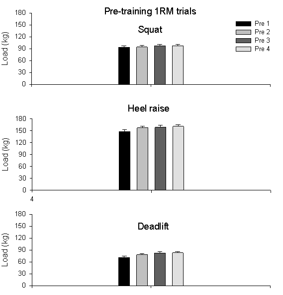
Figure 6. Pre-training 1RM values (mean ± SE) for 33 subjects, where Pre 1-4 are the four successive 1RM trials prior to training.
Subject Characteristics
An overview of the subject characteristics and the means for the groups are provided in Table 1. The groups are listed according to subject ID in Table 2. At the beginning of the study, each groups contained seven subjects.
|
Table 1. Subject baseline characteristics. All values are group mean ± SE. (N=7 for each group). |
|||
|
Group |
Age (yr.) |
Height (cm) |
Weight (kg) |
|
Control |
32.0 ± 2.1 |
179 ± 1.5 |
86.4 ± 2.6 |
|
FW |
33.0 ± 2.8 |
180 ± 3.3 |
77.3 ± 3.9 |
|
IRED3 |
32.0 ± 2.7 |
180 ± 3.0 |
81.2 ± 3.5 |
|
IRED6 |
35.0 ± 2.0 |
181 ± 3.4 |
83.1 ± 4.6 |
There are no differences (pre-training) in the baseline characteristics when comparing the four groups for age (P= 0.794), height (P=0.938) and weight (P=0.397).
|
Table 2. Overview of the groups with the subjects ID. (N=7 for each group) |
|||
|
Control |
FW |
IRED3 |
IRED6 |
|
14 |
3 |
2 |
1 |
|
16 |
8 |
4 |
6 |
|
17 |
10 |
11 |
9 |
|
30 |
17 |
18 |
24 |
|
34 |
20 |
19 |
25 |
|
35 |
22 |
21 |
26 |
|
36 |
23 |
28 |
29 |
Pre-training One Repetition Maximum (1RM)
1RM values for each subject were obtained in four sessions, one session per week, prior to the beginning of the study. Based on published data (McCarthy et al., 1994), we chose the mean of the highest two 1RM values obtained for each subject represent the 1RM value. When comparing the sessions, there were no differences in pre-training 1RM values for the squat exercise. However, the pre-1RM value for heel raise and deadlift increased over time (P<0.001), with the highest values appearing, on average, during the third and the fourth session. The pre-training 1RM values for each exercise are presented in Figure 6.

Figure 6. Pre-training 1RM values (mean
± SE) for 33
subjects, where Pre 1-4 are the four successive 1RM trials prior to training.
After calculating the pre-training1RM, the subjects were subsequently assigned to one of the four groups. The calculated pre-training 1RM value for each exercise is depicted per group in Figure 11. At the beginning of the training study, the means of the 1RM values did not differ between groups for squat (P=0.869), heel raise (P=0.642) and deadlift (P=0.862) exercises. The calculated mean values are from the 28 subjects (n=7 for each group) who completed 16-week training study. Subjects that did not finish the study are not represented.
Training Data
To represent changes in training intensity throughout the training program, three high-intensity days were chosen near the beginning, midway, and at the end of the program. The first time point (PRE) represents the first high-intensity session ( ~ session 10) of the fourth week of training, the second time point (MID) represents the first high-intensity session ( ~ session 28) after the mid-1RM testing, and the third time point (POST) represents the last high-intensity session ( ~ session 44) before the tapering in training intensities, which occurred one week before post-training 1RM testing ( ~ session 44). The peak forces used in the statistical analysis for the iRED training groups are the average of the peak forces recorded during training.
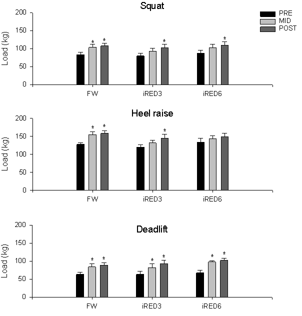
Figure 7. The intensities, expressed as load (kg),
for all the three exercises for the exercising groups at the representative
three time points. PRE represents the 10 th session, MID represents
the 28 th session and POST represents the 44 th session,
by approximation. Bars are group means ±
SE. Asterisk depicts statistical difference
from pre-training value.
At each of the three time points, the peak force, range of motion (ROM), and number of training repetitions (REPS) were compared. Figure 7 shows the peak forces for the iRED groups, as well as the training weight for the free weight group, for the three exercises.
The three time points are represented as PRE, MID and POST, and will be addressed as such in the statistical analysis. Due to hardware failure, one subject (9, iRED6) had his first high-intensity session at session 18, which was not a consistent time-point with the rest of the subjects. This subject was therefore excluded from the statistical analysis. Since FW data was recorded in load and iRED data was recorded in peak force no statistical analyses could be made to compare the FW group and the iRED groups.
Mean squat forces changed significantly throughout the training period for all exercising groups. Training intensity increased 25.6 ± 5.5% for the free weight group (P<0.001) over the first half of the study, and then reached a plateau. Training intensity increased 30.8 ± 4.7% after 16 weeks of training. Training intensity also increased in the iRED groups pre- to post- by 28.3 ± 3.5% (P= 0.003) and 28.8 ± 13.2% (P=0.008), for the iRED 3 and iRED6 group, respectively. There were no statistically significant differences between the iRED groups.
The training intensity of the heel raise for the FW group increased during the first half of the study from 21.2 ± 4.6% (P<0.001) to 24.0 ± 3.8, with no further changes observed during the second half. The intensity increased 20.5 ± 5.5% (P=0.007) for the iRED3 group by the last training period whereas the iRED6 did not show significant changes in training intensity for the heel raise.
Training intensities for the deadlift also increased over time for all groups. Increases were observed during the first half of the training study, and when comparing pre- to post-training. The increases for the first half were: 33.1 ± 8.1% (P<0.001), 32.3 ± 6.7% (P=0.002) and 50.4 ± 12.1% (P=0.001), for the FW, iRED3 and iRED6 group, respectively. No further changes were observed for the second half. At the end of the study, the increases in training intensity were: 41.8 ± 7.3% (P<0.001), 53.8 ± 11.9% (P=0.000) and 56.3 ± 10.8% (P=0.000) percent for the FW, iRED3 and the iRED6 group respectively. There were no differences in intensities between the iRED groups at any of the given time points for any of the exercises.
Range of Motion (ROM)
The ranges of motion recorded during the three representative days for each exercise verify that the exercise technique did not change during the study for each subject. The deflection of the squats, heel raises and deadlifts did not change over time for the groups and there were no differences over time between the groups (P>0.05). However, the average deflection for the deadlift was significantly higher for the FW when compared to the iRED3 group (P= 0.005) and the iRED6 group (P=0.015). Figure 8 depicts the average ROM for the three representative time points.
Repetitions (REPS)
Training volume, expressed as number of training repetitions, decreased 22.6 ± 4.1% over time for the FW group (P= 0.037) during the squat exercise. The number of post training repetitions at the MID time point was less for the FW group than the iRED3 group (P= 0.029), but there were no differences at the end of the training study. At all time points, the number of training repetitions performed by the iRED6 group were higher than for the 3-set groups (P=0.000). The total training volume for the three days was approximately 83% more than the FW group and 58% more than the iRED3 group. Figure 9 depicts the average REPS for the three representative time points.
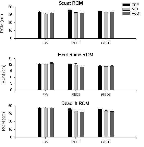
Figure 8. The range of motion
± SE. of each exercise, for each group. The
first time point represents the 10 th session, the second time
point represents the 28 th session and the last time point represents
the 44 th session, by approximation.
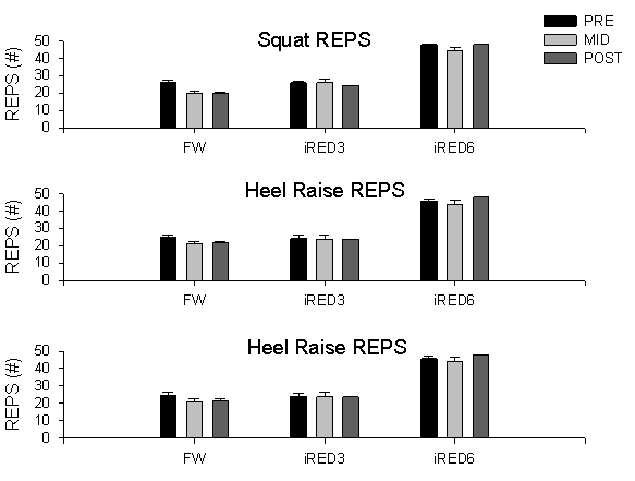
Figure 9. The number of training reps ±
SE. for each exercise for each group at the
representative three time points. The first time point represents the 10
th session, the second time point represents the 28 th session
and the last time point represents the 44 th session, by approximation.
Significant differences are marked with an asterisk.
Training Compliance
An overview of the training compliance for the exercising subjects is presented in table 3.
|
Table 3. Training compliance. The total amount of sessions is depicted in column three, where the 1RM sessions were counted as high-intensity training sessions. The average number of sessions per week is provided in the last column. Values are mean ± SE. |
|||
|
Group |
Training duration |
Sessions |
Sessions/week |
|
FW |
119.7 ± 4.5 |
48.4 ± 0.6 |
2.9 ± .01 |
|
iRED3 |
122.1 ± 4.0 |
49.7 ± 0.8 |
2.9 ± 0.1 |
|
iRED6 |
129.3 ± 4.4 |
47.4 ± 1.0 |
2.6 ± 0.1 |
During the training study, several of the subjects experienced injuries. The number of missed sessions and the nature of the injuries are presented in Table 4.
Table 4. Subject injury reports, with suspected cause and the total number of training days that the subjects did not train as a consequence of the injury.
|
Group |
Subject |
Injury |
Suspected cause |
Missed days |
|
iRED 6 |
1 |
GT Bursitus |
Over training |
14 |
|
iRED 6 |
9 |
Proc. Spin. C7 Fracture |
Misplacing HR Bar during 1RM |
7 |
|
iRED 3 |
12 |
Quadriceps Strain |
Flag Football injury |
5 |
|
iRED 6 |
13 |
L. Back Pain |
Over training |
7 |
|
FREE |
17 |
L. Back Pain |
Over training |
5 |
|
iRED 6 |
26 |
Patella Tendonitis |
Basketball |
14 |
|
iRED 6 |
29 |
Quadriceps Tendonitis |
Over training |
7 |
Hardware Failure, CRES, AUG and ROM
CRES Mode
Only the phase one groups training on the iRED experienced prolonged hardware failures. For several training sessions, the subjects trained with the contingency resistive exercise system (CRES). Phase one iRED3 subjects (2,4 and 11) resorted to the CRES system 5.7 ± 0.3 times, whereas the impact for the phase one iRED6 group was less, 2.3 ± 0.3 times. The second phase groups never trained with the CRES system. An overview is provided in Table 5.
|
Table 5. Overview of number of sessions in the CRES and AUG mode for each subject. Both iRED groups with their subjects (ID) for each phase. CRES= contingency resistive exercise system mode, AUG= augmentation mode, SQ=squat, HR= heel raise and DL deadlift. |
||||||||
|
|
ID |
CRES |
AUG |
|||||
|
SQ |
HR |
DL |
SQ |
HR |
DL |
|||
|
iRED3 |
Phase one |
2 4 11 |
6 5 6 |
6 5 6 |
6 5 6 |
3 -- 2 |
3 12 29 |
-- -- -- |
|
Phase two |
18 |
-- |
-- |
-- |
-- |
-- |
-- |
|
|
19 |
-- |
-- |
-- |
-- |
24 |
-- |
||
|
21 28 |
-- -- |
-- -- |
-- -- |
-- -- |
4 -- |
-- -- |
||
|
iRED6 |
Phase one |
1 |
2 -- |
2 -- |
2 -- |
-- 7 |
-- 34 |
-- 2 |
|
6 |
||||||||
|
Phase two |
24 25 26 29 |
-- -- -- -- |
-- -- -- -- |
-- -- -- -- |
-- -- -- -- |
29 5 16 17 |
-- -- -- -- |
|
AUG Mode
Because of the capacity limitations of the iRED, 6 subjects of both iRED groups trained on iRED in the augmentation mode. Since the Heel Raise exercise produces the highest absolute force values, the subjects used the augmentation mode most frequently with the heel raise exercise, 15.7 ± 9.9 and 16.3 ± 4.6 times by the iRED3 and iRED6 group respectively. Only three subjects (subjects 2, 11 and 6) used the AUG mode during the squat and one subject (subject 6) used the AUG mode for the deadlift exercise (2 times).
Range of Motion
When comparing the range of motion between the groups, no differences were observed. However, the range of motion was lower for the deadlift exercise for the iRED3 group (P=0.014) and the iRED6 group (P=0.001) when comparing their pre-training deadlift 1RM ROM to the average training ROM. Data is provided in Figure 11.
Mid-, and Post-training 1RM Sessions
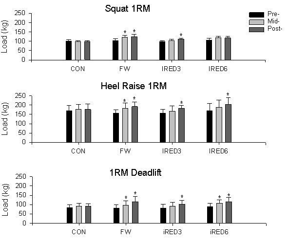
Figure 10. Pre-, mid- and post-training 1RM values
± SE
for the three exercises for all
the groups. Significant differences are marked with an asterisk
Halfway trough the training study ( ~ 72.7 ± 11.4 days after the last pre-training 1RM) mid-training 1RM values were obtained. Post-training 1RM values were obtained after completion of all the training sessions, which was 132.8 ± 12.8 days after the pre 1RM. Figure 10 shows the averaged pre-, mid- and post-training 1RM values for the groups for the different exercises.
Mean squat 1RM values increased over time for the FW group and for the iRED3 group. The mean pre-training 1RM value for the FW group was significantly different from mid- (17.1 ± 6.4%) and post-training (20.1 ± 5.7%) 1RM values (P=0.003, and P=0.000, respectively), but there was no difference between the mean mid- and post-training values. For the iRED3 group, the 1RM value increases from pre- to post-training with 15.8 ± 3.7% (P=0.015). For both the Control and the iRED6 group, there were no differences over time. During the study, there were no differences between groups at any of the given three time points.
Mean Heel Raise 1RM values increased over time for all the exercising groups. The mean pre-training 1RM value for the FW group increased 17.4 ± 4.8% during the first half (P=0.000), with a 22.9 ± 2.8% total increase. For the iRED3 group, the pre-training value increased by 16.9 ± 2.6% (P=0.001), and the iRED 6 group increased in strength 23.0 ± 6.6% (P=0.000), when comparing the pre-training values to the post-training values, with no changes between the mean mid- and post-training 1RM values. During the study, there were no differences between groups at any of the given three time points.
Mean deadlift 1RM values also increased over time for all the exercising groups. The mean pre-training 1RM value for the FW group increased between all the three time points; P-values are: 0.004, 0.000 and 0.003 for increases between pre- to mid- (20.4 ± 3.9%), mid- to post- (17.6 ± 3.7%) and pre- post-training (41.8 ± 6.9%) values. For the iRED3 group, the post-training value increased (25.1 ± 5.0%) in relation to the pre-training 1RM value, without a difference between the mean mid- and post-training 1RM values (P=0.000). The mean iRED6 group 1RM value increased by 21.7 ± 6.7% (P=0.009) and 30.5 ± 7.3% (P=0.000, respectively, when compared to the mid- and post-training value, without a difference between mean mid- and post-training values. The control group showed no changes in strength over time. During the study, there were no differences between groups at any of the given three time points.
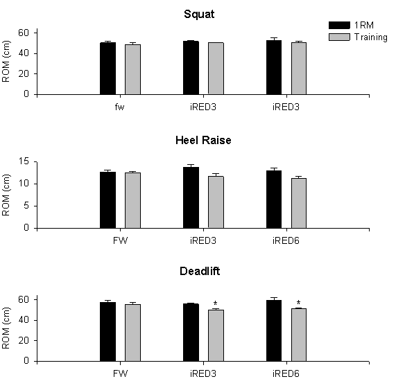
Figure 11. Comparison between the ROM ±
SE during the 1RM sessions and the average
ROM ±
SE during the training sessions. Significant differences are marked with an
asterisk.
There are no differences within and between the groups in comparing the range of motion during 1RM testing and deflection during squat training (P>0.05). However, differences are shown for the Heel Raise and deadlift exercise within the iRED training groups, where the average ROM during the heel raise training sessions was significantly lower than during the 1RM sessions (P<0.05). The ranges of motion for the FW group did not decrease.
Bone Mineral Content and Density
The DEXA scans reveal that there are no changes in bone mineral content (BMC) in the lower part of the spine (Lumbar L1 –L4) for any of the groups. For the FW group, there was an increase of 3.1 ± 1.1% (P=0.022) in L4 BMD. Figure 12 depicts the BMC and BMD values for L1 - L4 for each group. There are no changes in L4 bone area (P= 0.944). L4 BMC was 20.404 ± 0.442 vs. 20.359 ± 0.465g, pre to post.
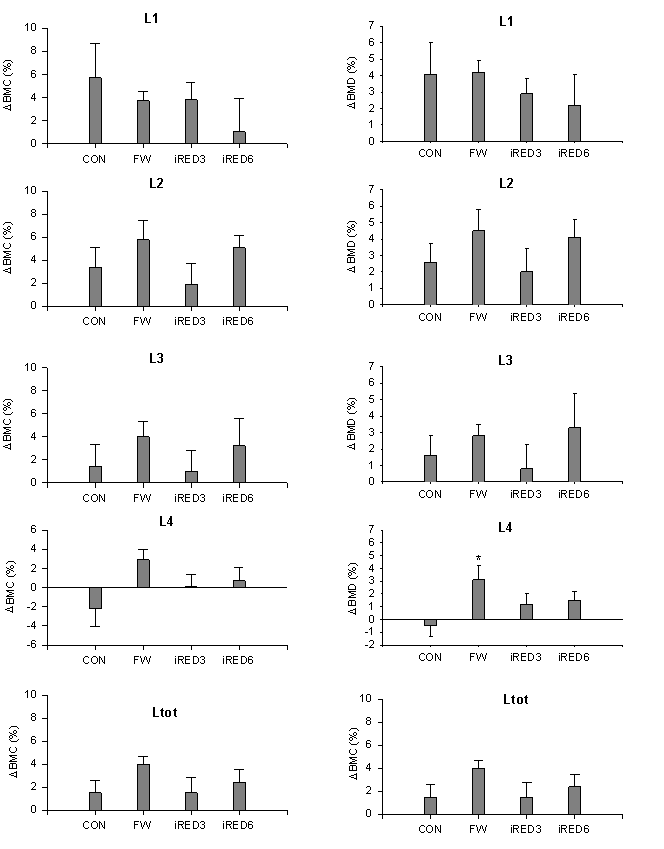
Figure 12. Dual energy X-ray absorptiometry scans
analyzed for the lumbar spine. On the left, the percent changes in bone mineral
content and on the right, the changes in bone mineral density. The bars represent
the mean changes for the groups ±
SE. Significant changes are marked with an asterisk.
The DEXA scans further revealed that there were no significant differences in BMC and BMD in the hip. We analyzed the DEXA scans for both BMC and BMD in the femoral neck, the trochanter, inter trochanter and total hip joint. Data is provided in Figure13.
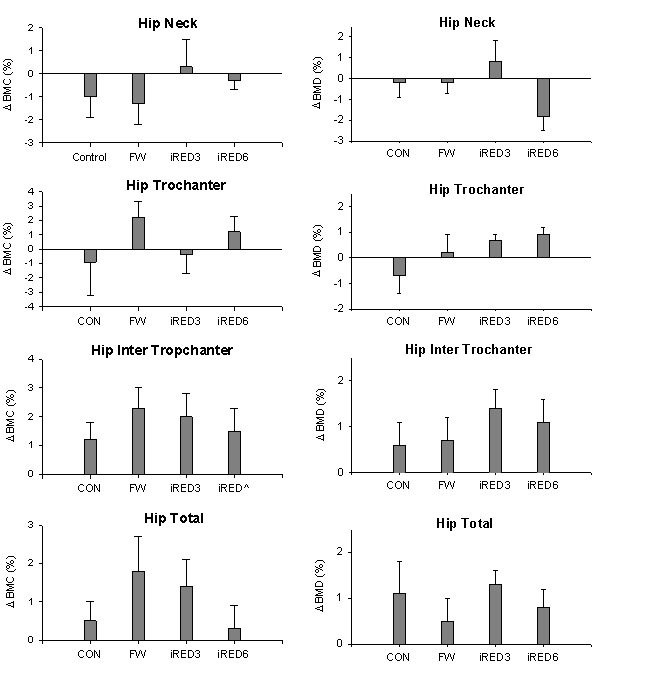
Figure 13. Dual energy X-ray absorptiometry scans
analyzed for the hip. On the left the mean percent changes in bone mineral content
and on the right the changes in bone mineral density. The bars represent the
mean changes for the groups ±
SE.
Whole body DEXA scans were analyzed for changes in BMD and BMC in the lower limbs. As shown in Figure 14, there were no changes in total leg BMC and BMD.
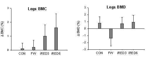
Figure 14. Dual energy X-ray absorptiometry scans
analyzed for the legs, with on the left the mean percent changes in bone mineral
content and on the right the changes in bone mineral density. The bars represent
the mean changes for the groups ±
SE.
Whole Body DEXA scans were analyzed for differences in total leg bone mineral content (BMC) and total leg bone mineral density.
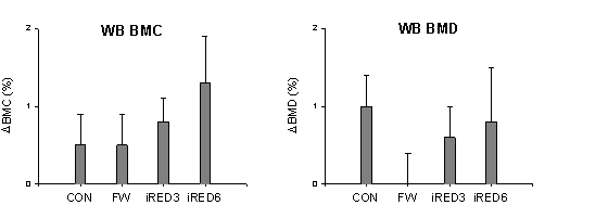
Figure 15. Dual energy X-ray absorptiometry scans
analyzed for whole body (WB). On the left, the percent changes in bone mineral
content and on the right, the changes in bone mineral density. The bars represent
the mean changes for the groups ±
SE. Significant differences are marked with an asterisk.
Whole body DEXA scans were analyzed for changes in body composition (total mass, total lean tissue, total fat and total percent fat). The analysis was done for both the lower limbs and whole body.
For the lower limbs, lean (muscle) tissue increased 5.4 ± 1.2% over time for the FW group (P=0.000), 3.1 ± 0.5% for the iRED3 group (P=0.030) and 4.3 ± 0.6% for the iRED6 group (P= 0.001). Total leg mass, which consists of BMC, fatty tissue, and lean tissue, increased for the FW group 3.6 ± 0.9% (P=0.005), 3.3 ± 0.8% for the iRED3 (P=0.011) and 3.6 ± 0.8% for the iRED6 group (P=0.006). The changes in body composition observed in the legs are not accompanied by changes in whole body composition, except for the whole body lean tissue for the free weights, which increased 3.0 ± 1.1% (P=0.016). Figure 16 provides the data.
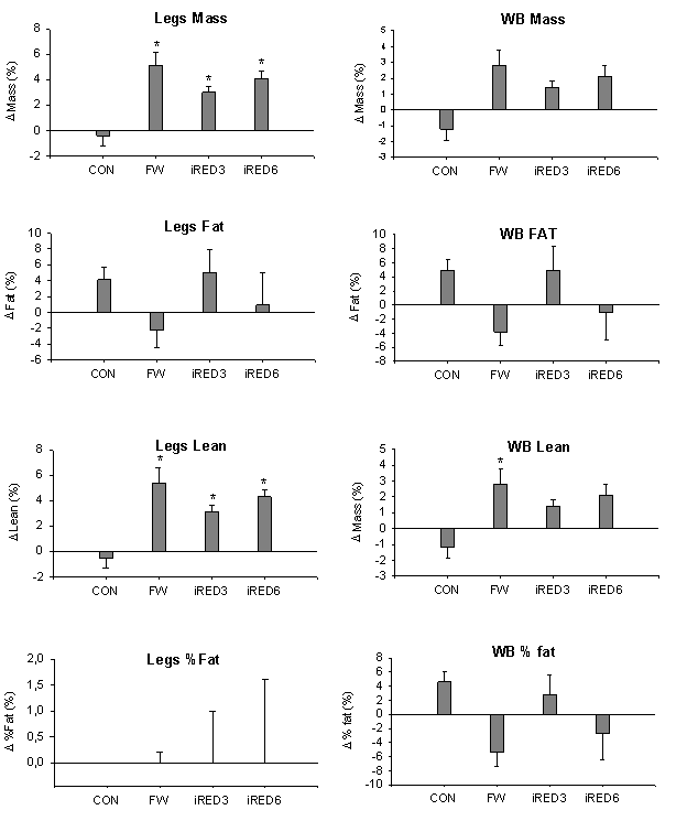
Figure 16. Dual energy X-ray absorptiometry scans
analyzed for body composition in legs and whole body. On the left, the percent
changes in body composition in the legs and on the right, the changes in body
composition of the whole body. The bars represent the mean changes for the groups
± SE.
Significant differences are marked with an asterisk.
Magnetic Resonance Imaging
Pre- and post-training muscle volumes, expressed as muscle pixels were determined using MRI. Table 6 shows the average percent change when comparing pre-training thigh and calf muscle volumes to post-training values. Two subjects (1 and 8) are excluded from the MRI data analysis, because of missing values. Due to hardware issues of the MRI system, the provided data set is incomplete and no statistical analysis was performed. The preliminary data, however, is provided in Table 6.
|
Table 6. Changes in muscle volume (mean ± SE), expressed as percent change in the thigh and the calf region. Con (n=3), FW (n=6), iRED3 (n=6), iRED6 (n=4). |
||
|
Group |
D Thigh (%) |
D Calf (%) |
|
CON |
1.2 ± 1.4 |
2.6 ± .85 |
|
FW |
9.2 ± 1.3 |
4.22 ± 1.0 |
|
IRED3 |
4.7 ± 1.1 |
6.1 ± 0.8 |
|
IRED6 |
6.7 ± 2.3 |
5.9 ± 1.7 |
Preliminary data suggests that muscle volume increased in all the exercising groups. For all the exercising groups, all subjects showed increases in thigh and calf muscle volumes.